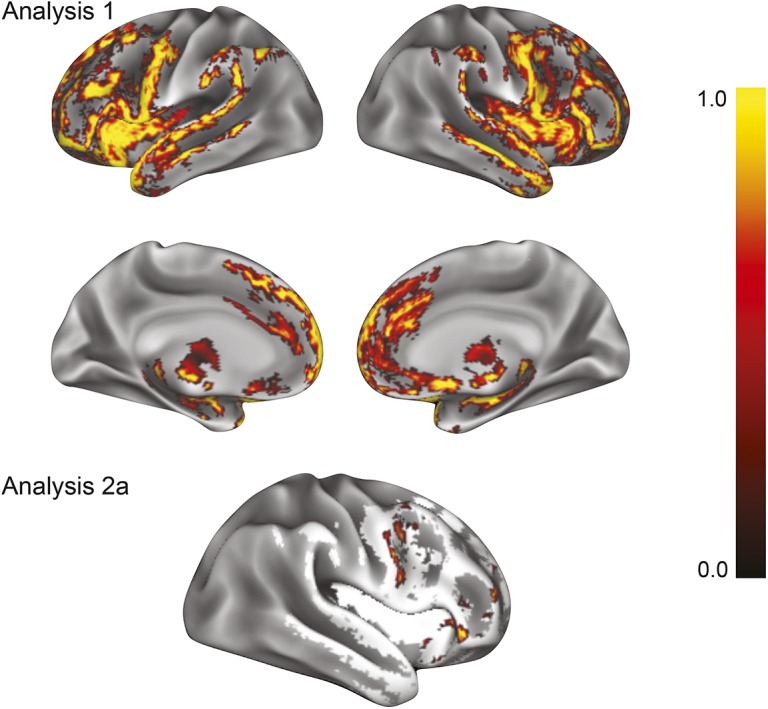
Figure 1. Higher cognitive reserve (CR) in patients with frontotemporal lobar degeneration (FTLD) and higher gray matter density (GMD) in diseased frontal lobe regions.

Analysis 1: Results of a nonparametric t test show regions of reduced GMD in patients with FTLD (n = 55) relative to demographically comparable controls (n = 90). Analysis 2a: Results of a regression analysis in patients with FTLD (n = 55) restricted to regions of reduced GMD from analysis 1 (white regions) demonstrate that higher GMD in the right dorsolateral prefrontal cortex, rostral frontal cortex, orbital frontal cortex, and inferior frontal gyrus is associated with higher CR index. Color bar represents 1 − p value with yellow representing highest significance.
