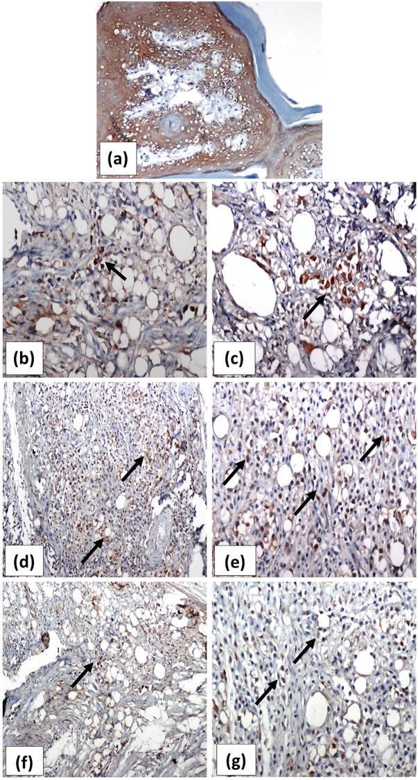
Fig 8.
Representative photomicrographs immunohistochemically stained sections of rats’ right hind paws with the anti-foxp3 antibody showing normal control rat ‘s paw in (a) with no inflammatory infiltrate and absent foxp3+ cells (x100). Scattered foxp3+ cells (↑) with brown stained nuclei of untreated arthritis group are shown in (b) and (c) (x200 and x400, respectively). (d) and (e) show marked increase in the foxp3+ cells in joints of ASMA-treated arthritis rat (x200 and x400, respectively). Moderate increase in the number of fox p3+ cells after treatment with ATSA is shown in f (x200) with its high power magnification in (g) (x400). ASMA; Autoclaved Schistosoma Mansoni antigen, ATSA; Autoclaved Trichinella Spiralis antigen.

