
Figure 1.
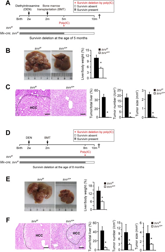
Survivin is essential in HCC development. (A) Experimental design for the DEN‐induced HCC model. Mx‐cre; svv f/f and svv f/f control mice were treated with DEN at 2 weeks of age and γ‐irradiated, followed by transplantation with bone marrows from svv f/f mice at the age of 2 months. No HCCs were detectable at the time of bone marrow transplantation. Five‐month‐old Mx‐cre; svv f/f and control mice were injected with poly(IC) intraperitoneally to induce liver‐specific Survivin gene depletion. Mx‐cre; svv f/f mice were referred to as svv Δli* after poly(IC)‐induced depletion of Survivin. All mice were sacrificed for HCC analysis at the age of 10 months. (B,C) DEN‐induced HCCs were analyzed in svv Δli* and svv f/f mice 5 months after poly(IC) treatment. (B) Liver/body weight ratios were measured. n = 5 for svv f/f mice; n = 6 for svv Δli* mice. (C) Hematoxylin and eosin (H&E) staining of liver sections. HCC was encircled with a dashed line. HCCs were quantified. n = 5 for svv f/f mice; n = 5 for svv Δli* mice. (D) Similar treatment as described in (A) was applied to Mx‐cre; svv f/f and svv f/f mice. However, Survivin was depleted with poly(IC) in Mx‐cre; svv f/f mice at the age of 8 months. (E,F) DEN‐induced HCC was analyzed in svv Δli* mice at the age of 10 months. (E) Liver/body weight ratios were measured. n = 7 for svv f/f mice; n = 5 for svv Δli* mice. (F) H&E staining of liver sections. HCCs were encircled with dashed lines, and HCCs were quantified. n = 5 for svv f/f mice; n = 5 for svv Δli* mice. Scale bars, 100 μm. * P < 0.05, t test.