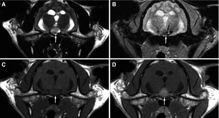
Figure 1.

Axial MRI images of the brain show a large nodule that is hyperintense on T2W images (A, white arrow), mildly hypointense on T1W images (C, white arrow), and strongly uniformly contrast enhancing (D, white arrow) in the region of the pituitary gland. A T2* gradient recalled echo sequence shows patchy regions of indistinct hypointensity (B, white arrow) with a small region of marked hypointensity or signal void (B, black arrow).
