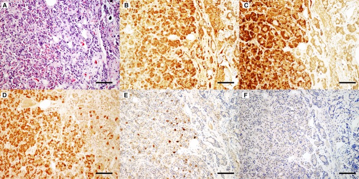
Figure 2.

Neoplastic cells vary in their intensity of eosinophilic cytoplasmic staining with many cells resembling acidophils (A). Hematoxylin eosin staining, bar = 200 μm. Neoplastic cells have diffuse cytoplasmic labeling for growth hormone (B), adrenocorticotropic hormone (C), and follicle‐stimulating hormone (D). Small numbers of cells within the neoplasm were positively labeled for melanocyte‐stimulating hormone (E), while labeling for thyroid‐stimulating hormone (F) was not observed. DAB, hematoxylin counterstain, bar = 100 μm.
