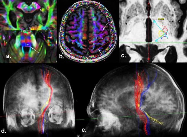
Figure 1.

The methodology of tracking the pyramidal tract (PT), the medial lemniscus (ML), and the dentate‐rubro‐thalamic tract (DRT). a and b: The regions of interest (ROIs) were placed at the cerebral peduncle and primary motor cortex for tracking the PT. Similarly, ROIs were placed at the dorsal brain stem and sensory cortex for visualizing ML. c: To define the lateral and posterior borders of tractography‐based–ventral intermediate nucleus (VIM), we created 2 lines at the medial border of the PT (red) and the anterior border of the ML (blue) at the anterior commissural‐posterior commissural level. We then generated a tractography‐based‐VIM ROI (a 3‐mm circle was first drawn at the angle of the PT and ML, and the thalamic ROI was then placed with its center coinciding with the center of the circle). The projections of this ROI were visualized, keeping the tracking angle constant at 60°. d: Coronal view of the 3‐dimensional reconstruction (semitransparent scalp and skull), demonstrating the relationship between the PT (red), ML (blue), and DRT and thalamo‐cortical projections from the VIM (yellow). The bilateral cerebellar contribution to the DRT can also be visualized. e: Sagittal view of the 3‐dimensional reconstruction (semitransparent scalp and skull), demonstrating the motor cortical projections from the VIM ROI (yellow) anterior to the ML (blue).
