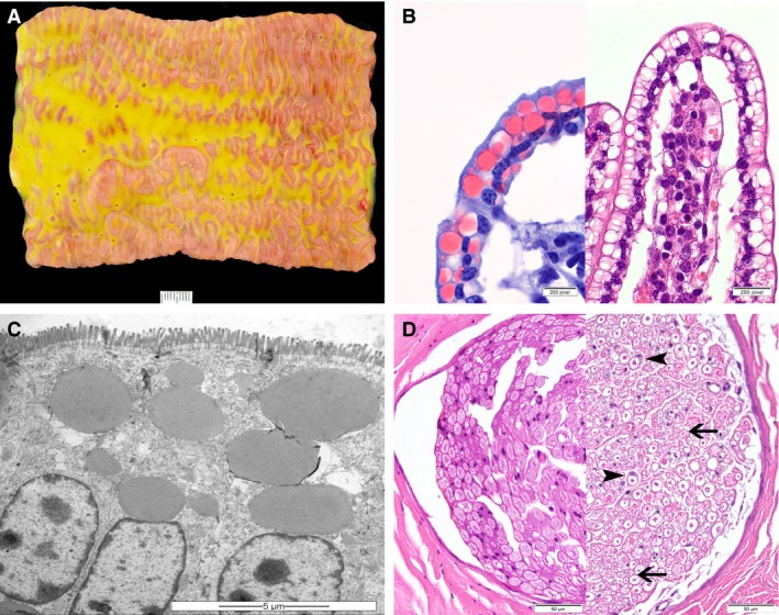
Figure 2.

Lesions of congenital cholesterol deficiency in Holstein cattle. (A) Macroscopic appearance of the small intestine and its content of the heifer no. 6. The intestinal content is foamy and greasy, varying from light‐yellow to green in color, and the mucosa is edematous (scale bar = 1 cm). (B) In routinely processed histological sections of jejunum, many enterocytes contain optically empty cytoplasmic vacuoles, which are shown to represent lipid inclusions in Sudan‐stained frozen sections (left) (100×). (C) Electron micrograph of enterocytes containing fat vacuoles within the apical cytoplasm (scale bar = 5 μm). (D) Sciatic nerve of the heifer (no. 6) (right), showing thin and irregular myelin sheaths (arrow) with Schwann cell activation (arrowheads) in comparison to a control animal of the same age (left) (HE, 40×).
