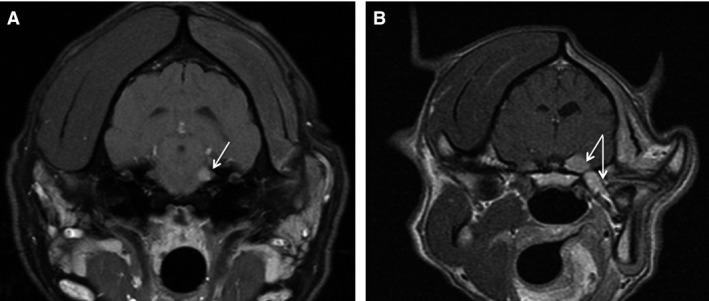
Figure 3.

Transverse MRI images of the brain. (A) Dog #7 presented with moderate muscle atrophy. Fat‐saturated contrast T1 MRI images revealed a small extra‐axial lesion in the region of the left trigeminal nerve. After treatment, the mass was no longer visible on MRI (not shown). (B) Dog # 1 presented with severe muscle atrophy. Contrast‐enhanced T1 images MRI revealed a left extra‐axial contrast‐enhancing mass with involvement of the extracranial trigeminal nerve extending to the left mandibular branch. The dog was euthanized at 855 days after seizure and diagnosis of sarcoma in the mouth that was possible progression.
