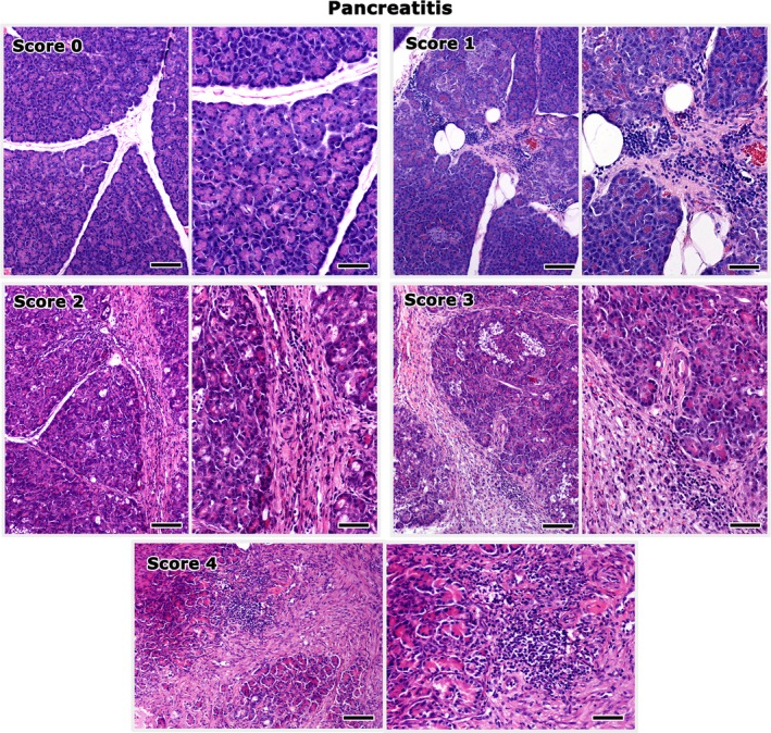
Figure 2.

Histopathological grading of pancreatitis lesions in cats. Score 0 corresponds to the normal histomorphology of the pancreas. Inflammatory cells are gathered mainly at the interlobular spaces. Interlobular fibrosis and replacement of the exocrine pancreas by connective tissue progressively increase in severity from score 1 to 4. Right panel images highlight areas from the images shown on the left at a higher magnification. Bars of left panel images: 100 μm. Bars of right panel images: 50 μm. Hematoxylin‐eosin.
