
Figure 2.
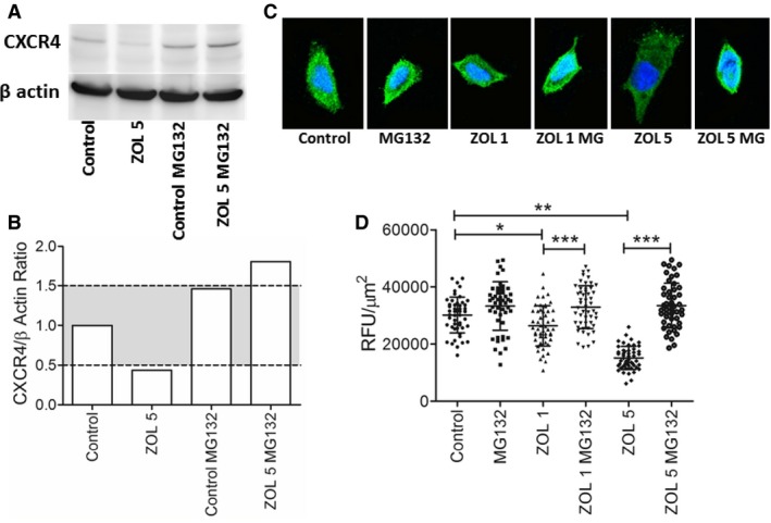
Qualitative modulation of CXCR4 protein expression by zoledronate and proteasome inhibition represented by (A) western blot analysis and (B) normalized graphical presentation where gray shaded region denotes ± 50% change in CXCR4 expression relative to untreated control cells. (C) Visual reduction in CXCR4 fluorescent intensities in K003 cells exposed to low concentrations of zoledronate, and complete normalization of CXCR4 expression with the co‐addition of MG132, a proteasome inhibitor. (D) Based upon a 50 cell count, quantitative changes in CXCR4 expression in K003 cells exposed to zoledronate with or without co‐addition of MG132, a proteasome inhibitor. Data expressed as mean ± SD and significance defined as *P < .05, **P < .01, and ***P < .001.