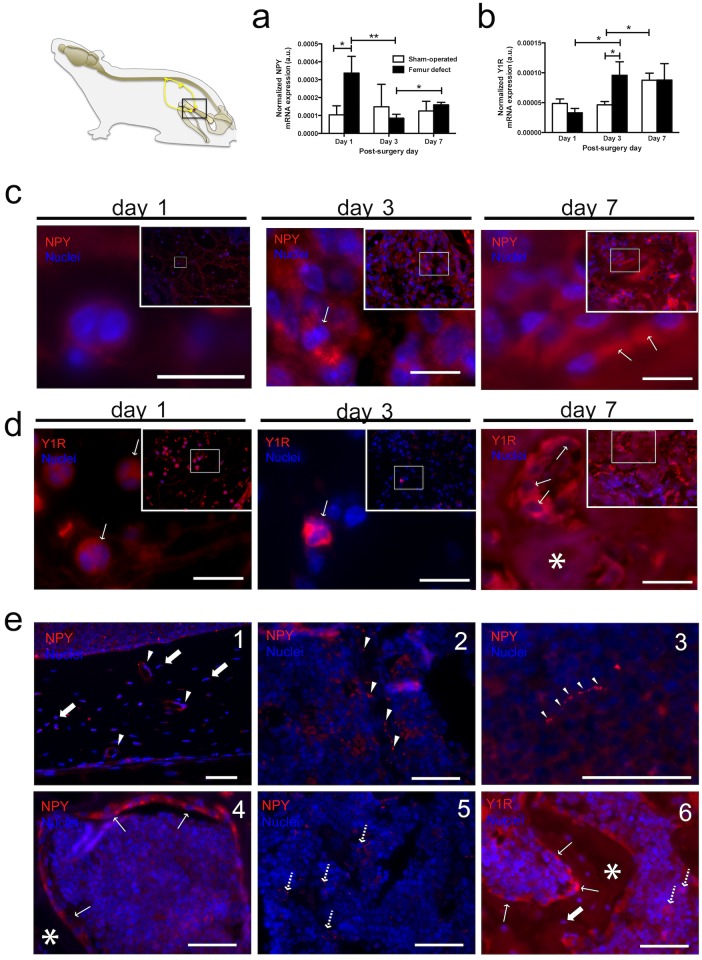
Fig 2. NPY system within bone microenvironment is responsive to bone defect.
NPY (a) and Y1R (b) mRNA expression levels were assessed at days 1, 3 and 7 post-surgery in femurs from femur-defect and sham-operated mice. (a) Femur-defect mice displayed 2.5-fold higher NPY mRNA expression levels at day 1 and (b) 2-fold higher Y1R mRNA expression levels at day 3 as compared to sham-operated animals. In (a) and (b) each column represents the mean + SEM, for 8 animals per group. *p<0.050; ** p<0.010. NPY (c) and Y1R (d) immunoreactivity were assessed in femoral histological sections from femur-defect and sham-operated mice. Within the defect, polymorphonuclear cells stained for NPY (c) and Y1R (d) were observed at day 1 post-defect. NPY-positive and Y1R-positive cells were also observed within the granulation tissue at day 3. At day 7 a high number of NPY- and Y1R-positive osteoblasts (c and d, respectively) were observed surrounding the newly formed bone. Panel (e) shows NPY and Y1R immunoreactivity in the areas adjacent to the defect. NPY-positive nerve fibers were observed alongside blood vessels in the bone (1; white arrowhead) and in the bone marrow (2; white arrowhead), and also scattered in the bone marrow (3; white arrowhead); NPY and Y1R immunoreactivity was also observed for osteoblasts (4 and 6, respectively; thin white arrow), osteocytes (1 and 6, respectively; thick white arrow) and bone marrow cells (5 and 6, respectively; dashed arrow). * indicates bone tissue. Panel (c) and (d)- scale bar = 10 μm; Panel (e)- scale bar = 50 μm.

