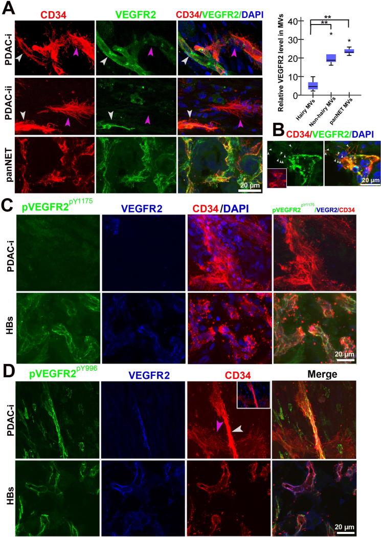
Figure 4. Low VEGFR2 expression and phosphorylation in microvessels that possess basal microvilli.
(A) VEGFR2 expression patterns in PDAC “hairy” or “non-hairy”microvessels and PanNET microvessels and endothelial tip cells (PDAC: yellow arrows, “non-hairy” microvessel; pink arrow, hairy microvessels; PanNET: small white arrows, VEGFR2 positive dots on filopodia). Quantitative analysis comparing VEGFR2 levels in “hairy” microvessels with “non-hairy”microvessels of PDAC and microvessels of PanNET. Statistical significance was assessed by student t test. (B) VERGR2 expression patterns in terminal endothelial cells of the PanNET microvasculature (white arrows, the VEGFR2 positive dots on filopodia). (C) Phospho-VEGFR2Y1175 (pVEGFR2Y1175) levels in “hairy” microvessels of PDAC and microvasculature in HBs. (D) Phospho-VEGFR2Y996 (pVEGFR2Y996) levels in “hairy”or “non-hairy” microvessels of PDAC and microvasculature in HBs (white arrow, “non-hairy” microvessels; pink arrow, “hairy” microvessels). See Movie S3.

