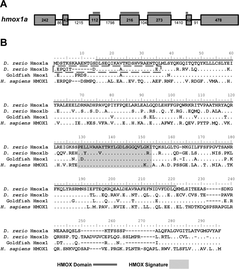
Figure 1. Zebrafish Hmox1a and Hmox1b contain conserved heme oxygenase domains and heme signature motifs.
A) Schematic of the zebrafish hmox1a gene. Exons are denoted by boxes and introns are denoted by lines. The numbers represent the nucleotide length for each exon or intron. The heme oxygenase domain (HMOX Domain) spans exons 3–6 and is heighted by the dark grey line. B) Multiple sequence alignment of HO-1 proteins from fish (goldfish and zebrafish) and human. Sequence alignment was generated using Clustal W. Amino acids identical to zebrafish Hmox1a protein sequence are designated by dots. The HO Domain (grey line) and the HMOX Signature (light grey box) are denoted within their respective sequences. The new N-terminal region of the Hmox1b protein sequence is denoted by a box with a dashed grey line. Zebrafish Hmox1a: NP_001120988.1; Zebrafish Hmox1b: KX664458; Human HMOX1: NP_002124.1; Goldfish (Carassius auratus) Hmox1 GenBank: AHI15729.1

