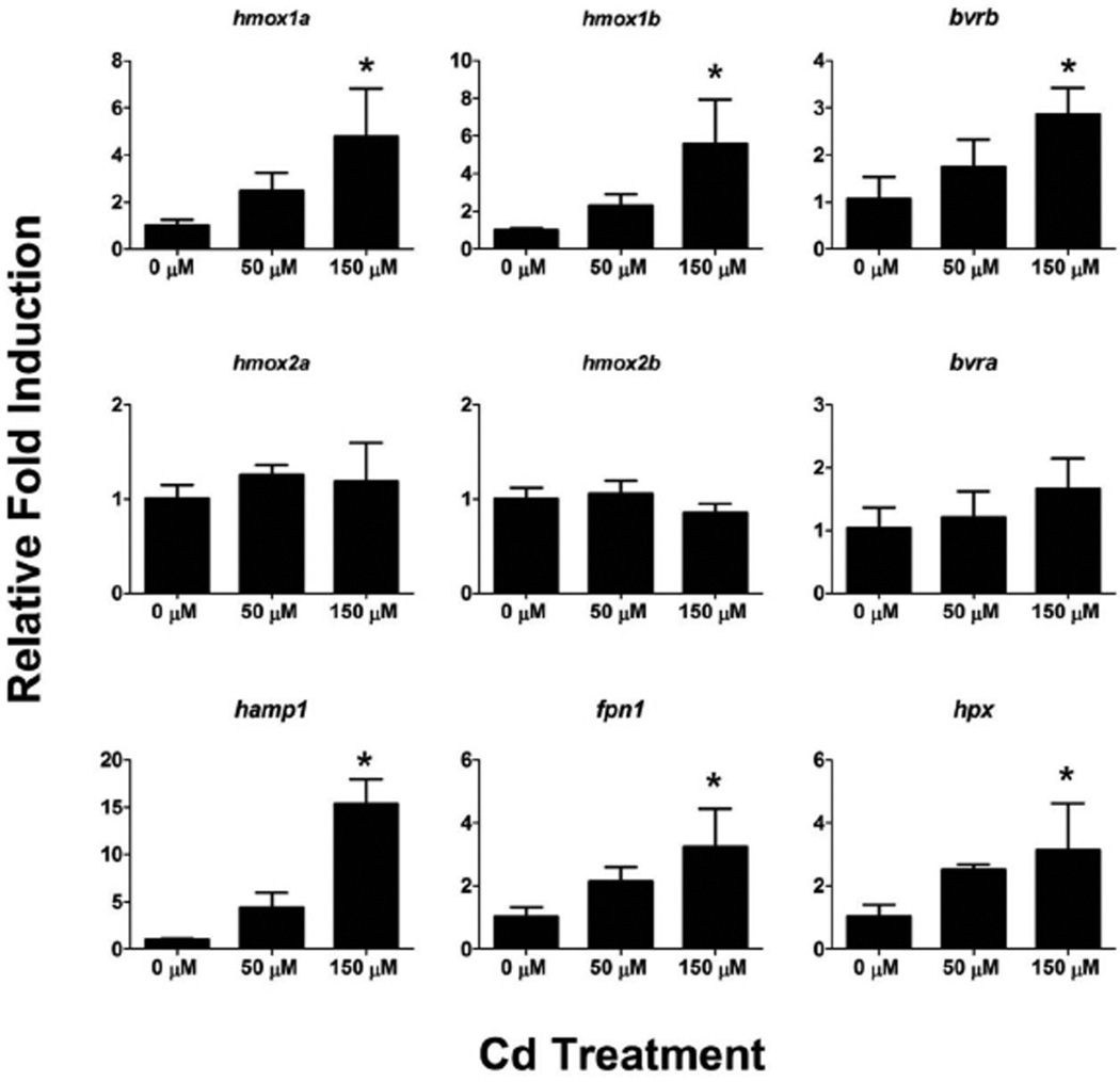
Figure 7. Effects of acute Cd exposure on gene expression in 76 hpf zebrafish.
Real-time RT-PCR was used to quantify changes in expression of heme degradation genes (hmox and bvr), as well as genes involved in heme and iron homeostasis, in whole zebrafish larvae at 76 hpf after a 4 hour exposure to Cd. Statistical significance in comparison to control embryos was determined using a one-way ANOVA followed by a Dunnett’s post hoc test (*p-value < 0.05). All values are normalized to 18S ribosomal RNA. Error bars represent one standard deviation; n = 3 biological replicates of 20 pooled embryos per treatment.

