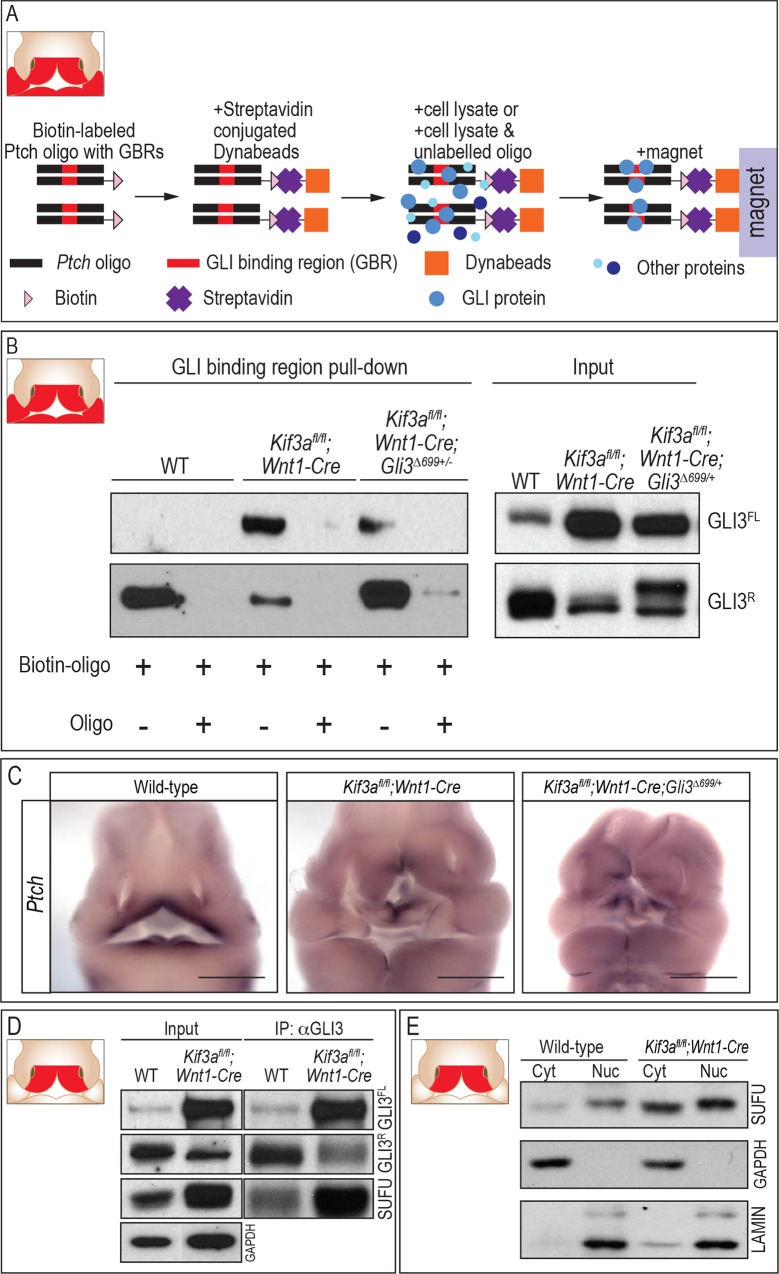
Fig 4. GLI isoforms binding to GLI binding regions is aberrant in ciliary mutants.
(A) Schematic diagram of experimental design. (B) Pull-down of GLI3 protein using Ptch oligo containing GBR (n = 3). (C) Whole mount in situ hybridization of Ptch in wild-type, Kif3afl/fl;Wnt1-Cre and Kif3afl/fl;Wnt1-Cre;Gli3Δ699/+ embryos. (D) SUFU pull down by GLI3 in the cytosolic fraction of FNPs from wild-type and Kif3afl/fl;Wnt1-Cre embryos. GAPDH was used as a loading control. (E) Nuclear fractionation of SUFU in wild-type and Kif3afl/fl;Wnt1-Cre FNPs. Lamin and GAPDH were used as loading control for nuclear and cytosolic fraction, respectively. Scale bars in C = 1000 μm. Inset schematics of facial prominences in A, B, D, E indicate FNP, maxillary prominence (MXP) and mandibular prominence (MNP) were harvested for experiments in A and B, while only the FNP (red) was harvested for experiments in D and E.

