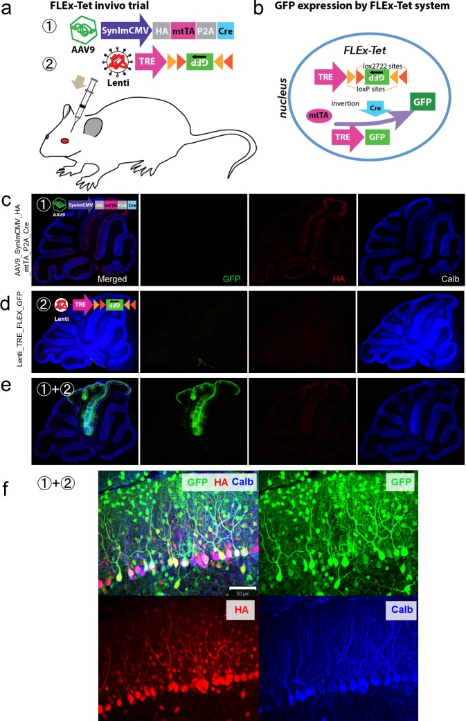
Fig 2. Validation of the FLEx-Tet system in mouse cerebella in vivo.
(a) The AAV9-SynImCMV-HA-mtTA-P2A-Cre vector bicistronically expressed HA-tagged mtTA and Cre recombinase under the control of a neuron-specific SynImCMV promoter (1). P2A, which is a ‘self-cleaving’ peptide sequence, was inserted between mtTA and Cre, whereas lentiviral vectors (Lenti-TRE-FLEx-GFP) carried TRE and an inverted GFP sequence flanked by loxP and lox2272 on both sides (2). AAV9 and/or lentiviral vectors were injected into 4-week-old mice. (b) Diagram depicting the FLEx-Tet system. Cre-mediated recombination and inversion of the GFP gene permitted expression of GFP protein in the presence of mtTA. (c-e) Immunohistochemistry of the mice that received an injection of the AAV9-SynImCMV-HA-mtTA-P2A-Cre vector (c), Lenti-TRE-FLEx-GFP vector (d) or the viral mixture (e). Two weeks after the viral injection, the mice were sacrificed, and the cerebellar slices were triple immunostained for GFP, HA and calbindin. Notably, GFP was expressed only in the cerebella of mice that received an injection of the viral mixture. (f) Magnified images of GFP-expressing lobules showing expression of GFP in various types of cortical neurons, including PCs, interneurons and granule cells. Scale bar, 50 μm.

