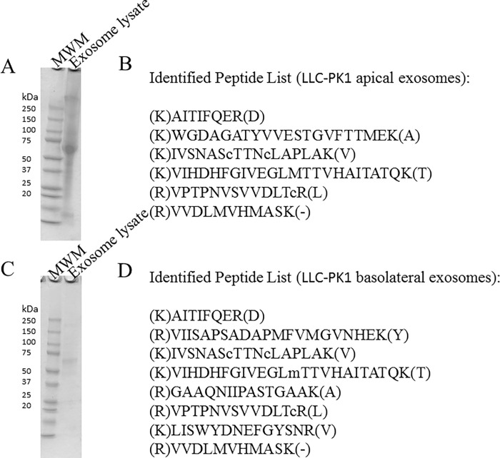
Fig 2.
Coomassie blue–stained sodium dodecyl sulphate-polyacrylamide gel electrophoresis analysis (A) and mass spectrometry analysis (B) of lysed exosomes isolated from conditioned media in the apical compartment of LLC-PK1 cells. Coomassie blue–stained sodium dodecyl sulphate-polyacrylamide gel electrophoresis analysis (C) and mass spectrometry analysis (D) of lysed exosomes isolated from conditioned media in the basolateral compartment of LLC-PK1 cells. Molecular weight markers (MWM) are shown in the first lane. Peptides listed in (B) and (D) are signature peptides corresponding to GAPDH.

