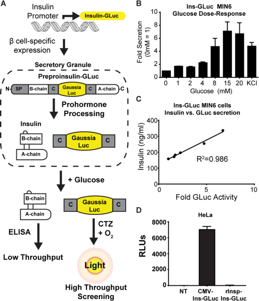
Figure 1.
A) Schematic for the creation and expression of the Ins-GLuc biosensor. B) Fold secreted luciferase activity in in MIN6 β cells expressing Ins-GLuc driven by the rat insulin promoter (rInsp). Raw RLUs were normalized to the activity at 0 mM glucose and expressed as fold ± SE. C) Correlation between GLuc luciferase activity and insulin secretion measured by ELISA from rInsp-Ins-GLuc-MIN6 subjected to a glucose dose-response curve (0, 1, 2, 4, 8, 20 mM). D) HeLa cells only express CMV-driven and not rInsp-driven reporter activity. Data are the average of 3 experiments ± SE.

