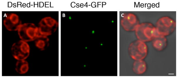
Figure 6. Visualization of clustered centromeres in P. pastoris by confocal microscopy.
This strain expressed DsRed-HDEL to label the endoplasmic reticulum in red. The ring visible in each cell is the nuclear envelope. In addition, the strain expressed Cse4-GFP to label centromeres in green. The merged image shows the two fluorescence signals overlaid on a transmitted light image of the cells. A cluster of centromeres is visible at the nuclear periphery in each cell. Scale bar, 2 μm.

