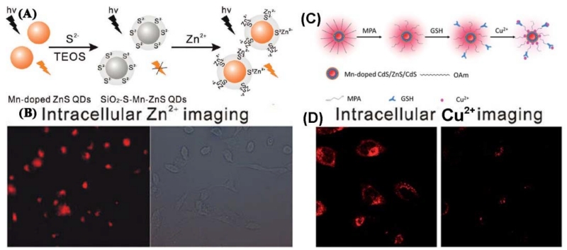
Figure 14.
(A) Schematic illustration for the fabrication of SiO2-S-Mn-ZnS QDs as a turn-on PL probe for Zn2+. (B) Intracellular imaging of Zn2+ with SiO2-S-Mn-ZnS QDs. HepG2 cells were incubated with Zn2+ in a fresh serum-containing medium for 20h. The collected Zn2+-uptaken HepG2 cells were then incubated with SiO2-S-Mn-ZnS QDs in a fresh serum-free medium for 4h and then in a fresh serum containing medium for 12h. (C) Schematic illustration for the formation of GSH-capped Mn-doped CdS/ZnS/CdS QD-based fluorescent nanosensor for Cu2+. (D). Confocal fluorescent images of HeLa cells before (left) and after (right) addition of Cu2+ (20 μM). (A and B: Adapted with permission from [237]. Copyright 2011 American Chemical Society, C and D: Adapted from [238] with permission of The Royal Society of Chemistry)

