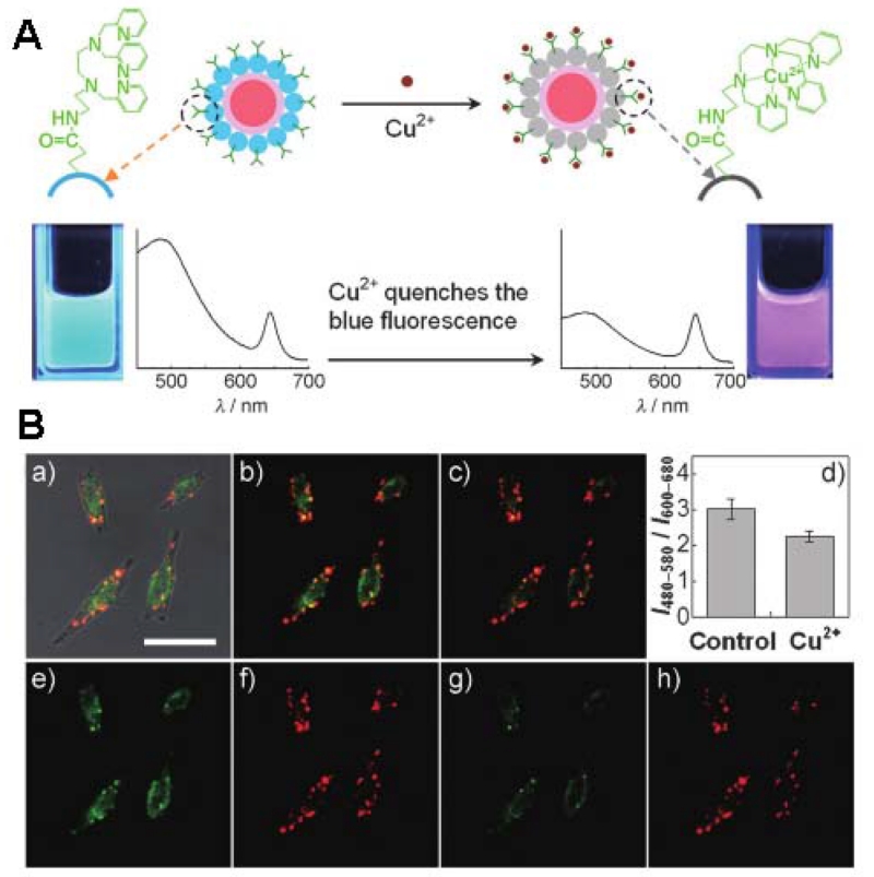
Figure 16.
(A) Schematic illustration of the dual-emission fluorescent sensing of Cu2+ based on CdSe@C-TPEA hybrid. (B) (a) The overlay of bright-field and fluorescence images of HeLa cells incubated with CdSe@C-TPEA. (b, c) Confocal fluorescence images of HeLa cells (b) before and (c) after the exogenous Cu2+ source treatment. (d) Bar graph representing the integrated intensity from 480 to 580 nm over the integrated fluorescence intensity from 600 to 680 nm; values are the mean ratio generated from the intensity from three randomly selected fields in both channel. (e, g) Confocal fluorescence images obtained from the 480-580 nm channel before and after the exogenous Cu2+ source treatment, while (f, h) are from the 600-680 channel. Scale Bar: 25 μm. (Reprinted with permission from [248]. Copyrights 2012 Wiley-VCH Verlag GmbH & Co. KGaA, Weinheim)

