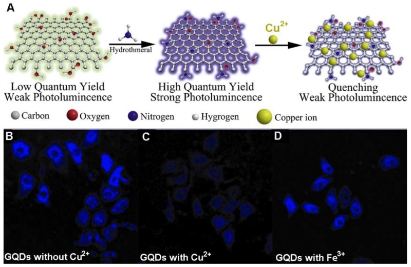
Figure 17.
Schematic representation of the preparation route for afGQDs, its quenching by copper ions, and intracellular Cu2+ profiling with afGQDs stained cells imaged without Cu2+ (B), with 10 μM Cu2+ (C), and with 10 μM Fe3+ (D) in dark field. (Reprinted with permission from [263]. Copyrights 2013 Wiley-VCH)

