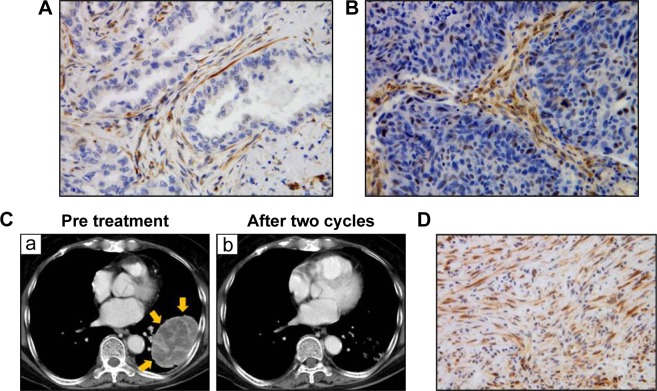
Figure 1.
Expression of SPARC in lung cancer tissues.
Notes: Immunohistochemical staining of lung adenocarcinoma tissue was performed using anti-SPARC antibody (A, ×400) and lung squamous cell carcinoma tissue (B, ×400). Counter staining was performed by hematoxylin. (C) (a) CT findings of the lung tumor in one case: a primary tumor was observed in the left lower lobe. The tumor position is indicated by the yellow arrows. After two cycles of chemotherapy, the tumor was markedly reduced (b). Immunohistochemical staining of TBLB samples from the primary lesion before treatment were performed using anti-SPARC antibody (D, ×400).
Abbreviations: SPARC, secreted protein acidic and rich in cysteine; CT, computed tomography; TBLB, transbronchial lung biopsy.

