. 2016 Sep 28;5:e19732. doi: 10.7554/eLife.19732

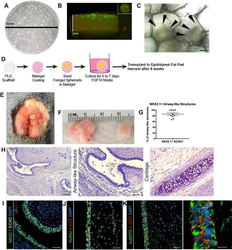
Figure 1. Transplanted HLO-scaffold constructs engrafted, grew and possessed airway-like structures.

(A) PLG scaffold are 5 mm in diameter with honeycomb-patterned architecture. (B) The majority of Di-O labeled 1d HLOs (green) remained at the surface of the scaffold with a few organoids descending toward the middle of the scaffold. Inset shows aerial view of the scaffold with 1d HLOs (green) scattered throughout. (C) 1d HLOs settled within the pores of the scaffold. Scale bar represents 100 µm. (D) PLG scaffolds were seeded with 1d HLOs and cultured for 5 to 7 days in vitro in media supplemented with FGF10. The HLO-laden scaffolds were then transplanted into the mouse epididymal fat pad and harvested at 8 weeks. (E) HLO-scaffold (dotted line) was placed in mouse epididymal fat pad. (F) Transplanted HLOs (tHLOs) ranged from 0.5 cm to 1.5 cm in length. (G) The average number airway-like structures that were NKX2.1+ ECAD+ out of all ECAD+ structures was 86.19% +/- 4.14% (N = 10, error bars represent SEM). (H) H&E of tHLOs showed airway-like structures (right two panels, low and high mag) and pockets of cartilage (left panel). Scale bar at low mag represents 200 µm and high mag 100 µm. (I) Airway-like structures outlined by ECAD (white) expressed the lung marker NKX2.1 (green). Scale bar represents 50 µm. (JK) Both the epithelium (β-CAT, red) and mesenchyme expressed the human nuclear marker, HUNU (J, green) and the human mitochondrial marker huMITO (K, green). Scale bars represent 50 µm in JK and 10 µm in high mag image in K.

DOI: http://dx.doi.org/10.7554/eLife.19732.003

Figure 1—source data 1. Summary of NKX2.1+ epithelial structures in individual tHLOs.
The number of ECAD+ structures were counted and scored as positive or negative for NKX2.1 expression. The percent NKX2.1+ epithelial structures were calculated (NKX2.1+ECAD+/total ECAD+) for each tHLO (conditions listed). The averages are listed in the bottom row.
DOI: 10.7554/eLife.19732.004

Figure 1.

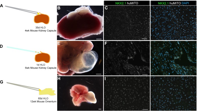
Figure 1—figure supplement 1. Highly vascular engraftment sites did not maintain lung epithelium.

Figure 1—figure supplement 1.

(A) 35d HLOs were transplanted under the mouse kidney capsule and retrieved after 4 weeks. (B, E, H) Whole-mount of the retrieved tissue within the kidney capsule. Transplanted tissue (white) was situated adjacent to the kidney (pink). (C) Tissue retrieved from the kidney capsule did not express the lung marker NKX2.1 (green), but expressed the human mitochondrial marker (huMITO, white) n = 6. (D) 1d HLOs were placed under the kidney capsule and retrieved after 6 weeks. (E) 1d HLOs transplanted under the kidney capsule consisted of transparent cysts and dense tissue. (F) Tissue retrieved from the kidney capsule did not express NKX2.1 (green), but expressed huMITO (white) n = 1. (G) 65d HLOs were sewn into the omentum surrounding the stomach and proximal intestine. The tissue was retrieved after 12 weeks. (H) The outgrowth (white) is the transplant after 12 weeks. (I) The majority of the tissue did not express NKX2.1 (green), but all the retrieved tissue expressed huMITO (white). n = 13 Scale bars in B,E,H represent 250 µm and scale bars in C,F,I represent 50 µm.
Figure 1—figure supplement 2. HLO omentum transplants maintained lung epithelium poorly.

Figure 1—figure supplement 2.

(A) 2 out of 13 HLO omentum transplants expressed the lung marker NKX2.1 (green) and all the transplant tissue expressed the human mitochondria marker (huMITO, white). (B) 2 out 13 transplants possessed airway-like structures that expressed the basal cell marker P63 (green) and the ciliated cell marker FOXJ1 (white). Scale bars represent in AB 50 µm.
Figure 1—figure supplement 3. 1d HLOs grown on a scaffold and transplanted into the mouse epididymal fat pad expressed lung markers when harvested at 4 weeks.

Figure 1—figure supplement 3.

(A) PLG scaffolds were coated with a mix of Matrigel and DMEM/F12. 1d HLOs were then seeded onto the scaffold in 100% Matrigel. The scaffolds were cultured for 5 to 7 days in vitro in media supplemented with FGF10. The scaffolds were retrieved after 4 weeks. (B) Whole-mount image of 1d HLOs seeded on the scaffold and cultured for 5 days in vitro. (C) Tissue retrieved after 4 weeks expressed the lung marker NKX2.1 (green) and huMITO (white) within the airway-like structures n = 4. Scale bars in B represent 500 µm and in C represent 50 µm.
Figure 1—figure supplement 4. Transplanted HLO-scaffold constructs retrieved 4 weeks post-transplantation possessed airway-like structures that expressed basal and ciliated cell markers.

Figure 1—figure supplement 4.

(A) 4 week HLO-scaffold transplants had airway-like structures that expressed the basal cell marker P63 (green) and the ciliated cell marker ACTTUB (red), but did not express the club cell marker CC10 (white). (B) Airway-like structures expressed P63 (green) and the ciliated cell marker FOXJ1 (white). β-Catenin (βCAT, red) outlines the epithelium n = 4. Scale bars represent in AB 50 µm.
Figure 1—figure supplement 5. 1d HLOs grown on PLG scaffolds in vitro maintained NKX2.1 expression but did not generate airway-like structures after 8 weeks.

Figure 1—figure supplement 5.

(A) Whole mount image of scaffold grown for 8 weeks in vitro. Organoid tissue remained localized to the scaffold. Scale bar represents 500 µm. (B) HLOs within the scaffold expressed the markers NKX2.1 (green) and huMITO (white), but did not form organized airway-like structures. Scale bars represent 50 µm.