. 2016 Nov 1;5:e19298. doi: 10.7554/eLife.19298

Figure 2. ERK2 and Cdk1 phosphorylate the xCPEB4 NTD.

(A, B) In vitro kinase assay of (A) recombinant FL xCPEB4 or (B) fragments (1, 2 and 3) with metaphase II oocyte extracts treated with specific kinase inhibitors (roscovitine, Cdk inhibitor; SL0101, p90Rsk inhibitor; BI-2536, Plk1 inhibitor; U0126, MEK inhibitor; and FR180204, ERK inhibitor). DMSO was used as a negative control. The percentage of phosphorylation compared to DMSO is indicated (autoradiography, 32P). A representative experiment from three independent biological replicates is shown. See also Figure 2—figure supplement 12. (C) Two-dimensional phosphopeptide maps of xCPEB4 fragments (1, 2 and 3) phosphorylated with metaphase II (MII) oocyte extracts or with recombinant ERK2 or Cdk1/cyclin B. Phosphopeptides were resolved by thin-layer electrophoresis (TLE) followed by thin-layer chromatography (TLC). Arrows indicate sample origin. Phosphopeptides detected in MII were numbered. Asterisks (*) indicate phosphopeptides generated with recombinant kinases not present in MII. A representative experiment from three independent biological replicates is shown. See also Figure 2—figure supplement 3. (D) Disorder tendency (PONDR VL-TX predictor) and mass spectrometry phosphorylation site identification of xCPEB4. Asterisks (*) indicate phosphosites identified with MII extracts. Bold letters indicate phosphosites identified with either ERK2 or Cdk1/cyclin B. Green indicates ERK2 phosphorylation sites, while purple indicates Cdk1 phosphorylation sites. Red lines represent large disordered regions. The xCPEB4 fragments used are outlined. See also Figure 2—figure supplement 4.

DOI: http://dx.doi.org/10.7554/eLife.19298.004

Figure 2.

Figure 2—figure supplement 1. Loading controls for in vitro kinase assays with inhibitors.

Figure 2—figure supplement 1.

(A) Loading control for Figure 2A, in a Coomassie blue–stained gel. (B) Loading control for Figure 2B, in a Coomassie blue–stained gel.
Figure 2—figure supplement 2. In vitro kinase assay with Cdk1 and ERK2 immunodepleted extracts.

Figure 2—figure supplement 2.

(A) In vitro kinase assay of recombinant xCPEB4 full-length protein (FL), using metaphase II oocyte extracts immunodepleted for Cdk1 or Cdk1/ERK2. IgG was used as negative control. The upper panel shows the autoradiography (32P), while the lower panel shows the loading control in a Coomassie blue-stained gel. (B) Western blot showing the efficiency of depletion of Cdk1 and ERK2. Tubulin was used as a loading control.
Figure 2—figure supplement 3. Two-dimensional phosphopeptide maps kinetic.

Figure 2—figure supplement 3.

Two-dimensional phosphopeptide maps of xCPEB4 fragments (1, 2 and 3) phosphorylated with oocytes extracts collected at indicated times (GVBD, germinal vesicle breakdown; MII, metaphase II). Phosphopeptides were resolved by thin-layer electrophoresis (TLE) followed by thin-layer chromatography (TLC). Arrows indicate sample origin. Phosphopeptides are numbered. Three independent biological replicates were performed, with equivalent findings each time.
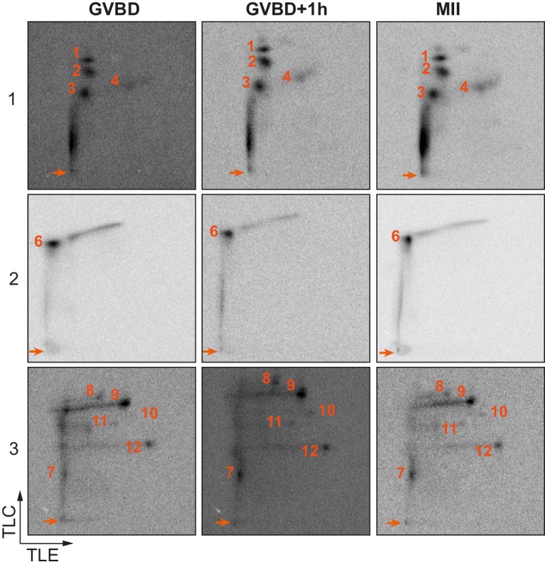
Figure 2—figure supplement 4. Confirmation of detected phosphosites.

Figure 2—figure supplement 4.

(A, B) Two-dimensional phosphopeptide maps of xCPEB4 fragments (1, 2 and 3), wild-type (WT) or phosphorylation mutants, phosphorylated with (A) metaphase II oocyte extract or (B) recombinant ERK2 or Cdk1/cyclin B. Phosphopeptides were solved by TLE followed by TLC. Arrows indicate sample origin. Phosphopeptides detected in WT conditions are numbered. Asterisks indicate unspecific phosphopeptides present only in xCPEB4 mutants. The mutations are specified at the bottom of panel A. (C) Lambda-phosphatase assay (λ) using extracts from oocytes overexpressing HA-CPEB4 collected at the indicated times (VI, prophase I; MII, metaphase II). Wild-type (WT) HA-CPEB4 and the 12A mutant (with 12 phosphorylation sites mutated to alanine) were analysed. The phosphorylation status of xCPEB4 was determined by western blot with anti-HA antibody and anti-tubulin (as a loading control). The control, C, corresponds to non-injected oocytes. Three independent biological replicates were performed, with equivalent findings each time.