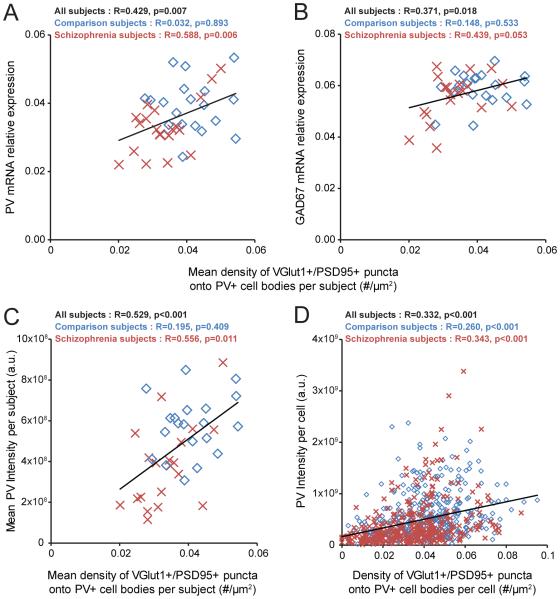
Figure 4.
Association between the density of excitatory synapses on PV+ neurons and activity-dependent expression levels of PV and GAD67 selectively in schizophrenia subjectsa
a In panels A and B, PV and GAD67 mRNA expression levels are plotted against the density of VGlut1+/PSD95+ puncta on PV+ cell bodies across subjects. The mean density of VGlut1+/PSD95+ puncta on PV+ cell bodies positively predicted the relative mRNA levels of PV (panel A) and GAD67 (panel B) in the schizophrenia subjects and not in the comparison subjects. In panels C and D, somal PV immunoreactivity levels are plotted against the density of VGlut1+/PSD95+ puncta on PV+ cell bodies across subjects or across all sampled PV+ neurons. Across subjects, the mean density of VGlut1+/PSD95+ puncta on PV+ cell bodies positively predicted the mean somal PV immunoreactivity levels in the schizophrenia subjects but not in the comparison subjects (panel C). Across all sampled PV+ neurons (N=725), the density of VGlut1+/PSD95+ puncta on PV+ cell bodies positively predicted somal PV immunoreactivity levels (panel D). Diamonds (blue): unaffected comparison subjects. Crosses (red): schizophrenia subjects. Trendlines (black): regression lines across all subjects.

