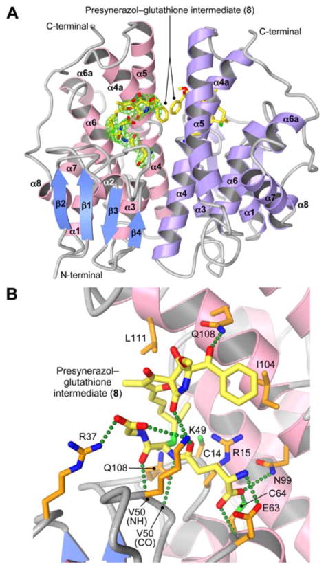
Figure 2.
Crystal structure of PsoE in complex with the GSH conjugate of presynerazol 8a. (A) Overall structure of PsoE (PDB ID: 5F8B) as a dimer. In the first PsoE molecule (left), α-helices and β-strands are colored in pink and blue, respectively, whereas the second molecule (right) is colored only in purple. Carbon atoms in the stick model of the bound ligand 8a are in yellow, whereas oxygen, nitrogen and sulfur atoms are in red, blue and green, respectively. Electron density for 8a in the first PsoE molecule (2Fo–Fc map contoured at 1.2 σ, green mesh) is shown. (B) The active site of PsoE, showing the interactions between 8a and PsoE. The protein side chain carbon atoms are in orange. Green dashed lines represent hydrogen bonds.

