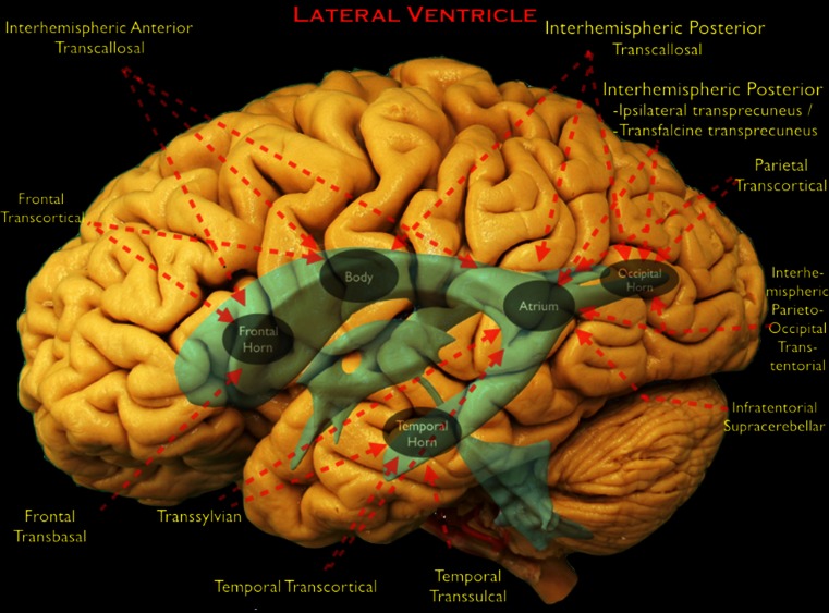
Fig. 1.
The surgical approaches to the lateral ventricle (LV) are shown on a lateral view of a cadaveric dissection of the brain. LV and third ventricle (TV) are shown in blue. Anatomical portions of the LV are depicted with gray ellipses. Red arrows show the direction of the approaches and the parts of the LV that can be reached by that individual approach

