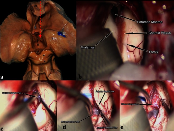
Fig. 5.
a The posterior part of the corpus callosum (CC) is removed, along with the posterior and superior walls of the LV, exposing the TV. The thalamus (T) forms the lateral walls of the posterior TV (III). The anatomical relation with the pineal gland (pi), superior colliculus (sc) and inferior colliculus (ic) can be seen. The red arrow shows the route leading to the FM and the TV through the CC. b An intra-operative picture demonstrating the anatomy of the choroidal fissure after entering to the LV. c Dissection between the fornix and the choroid plexus exposes the anterior septal vein. d The anterior septal vein and thalamostriate vein merge and form the internal cerebral vein. e Intraoperative picture revealing the velum interpositum (the roof of the TV) after retracting the venous structures and the choroid plexus

