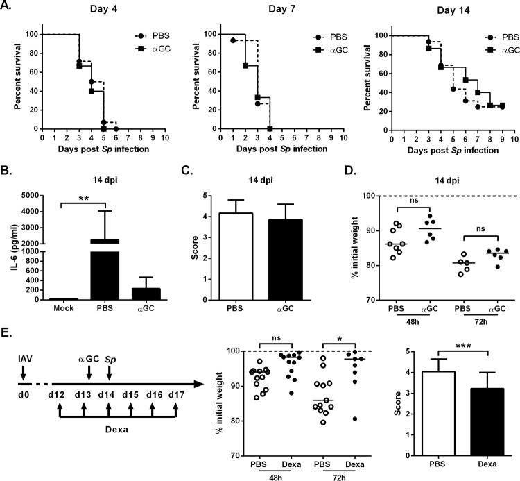
FIG 5 .
Effect of α-GalCer and dexamethasone treatment on the survival rate of superinfected animals. (A) IAV-infected mice were i.n. treated at different time points with PBS or α-GalCer (2 µg/mouse) (16 h before S. pneumoniae [Sp] challenge), and the survival of superinfected animals was monitored (n = 14 to 16/group, two pooled experiments). (B) IL-6 concentration in the serum of superinfected mice was quantified 24 h after S. pneumoniae infection. (C) For histopathologic examination, superinfected mice were killed 30 h after S. pneumoniae infection. Lung sections were scored blind for pneumonia with scores ranging from 0 to 5. (D) The body weights of the IAV-infected mice were measured 48 and 72 h after S. pneumoniae challenge and expressed relative to the weight at the time of bacterial challenge. (E) Overview of the procedure. IAV-infected mice were i.p. injected with dexamethasone (2.5 mg/kg) or vehicle 1 day before α-GalCer treatment (until day 17). Mice were challenged with S. pneumoniae at 14 dpi. (Middle panel) Modulations of body weights (relative to the weight before the bacterial challenge) are represented. (Right panel) Histopathological scores are indicated. (B, C, and E, right panel) Mean ± SD, n = 4 to 6 mice/group. One representative experiment out of two is shown. ns, not significant. *, P < 0.05; **, P < 0.01; *** P < 0.001 (one-way ANOVA Kruskal-Wallis test for panel B or Wilcoxon signed-rank test for panel E [right panel]).

