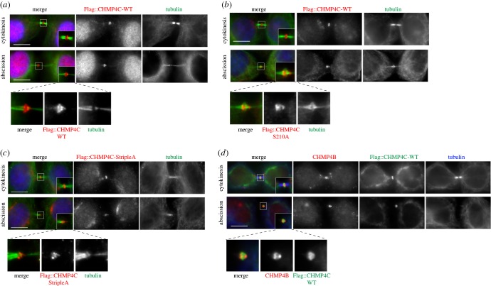
Figure 2.
Non-phosphorylatable forms of CHMP4C can form spiral structures at the abscission site. (a) HeLa Kyoto cells stably expressing Flag::CHMP4C WT were fixed and stained to detect Flag (red), tubulin (green) and DNA (blue). Insets show two time magnification of the midbody. A three times magnification of the midbody of the cell in abscission is shown at the bottom. Scale bars, 10 µm. (b) HeLa Kyoto cells stably expressing Flag::CHMP4C S201A were fixed and stained to detect Flag (red), tubulin (green) and DNA (blue). Insets show two times magnification of the midbody. A three times magnification of the midbody of the cell in abscission is shown at the bottom. Scale bars, 10 µm. (c) HeLa Kyoto cells stably expressing Flag::CHMP4C StripleA were fixed and stained to detect Flag (red), tubulin (green) and DNA (blue). Insets show two times magnification of the midbody. A three times magnification of the midbody of the cell in abscission is shown at the bottom. Scale bars, 10 µm. (d) HeLa Kyoto cells stably expressing Flag::CHMP4C WT were fixed and stained to detect Flag (green), CHMP4B (red) and tubulin (blue). Insets show two times magnification of the midbody. A three times magnification of the midbody of the cell in abscission is shown at the bottom. Scale bars, 10 µm. In all experiments, the shape and thickness of microtubule bundles were used as criteria to stage telophase cells as described [1,3].

