Figure 3.
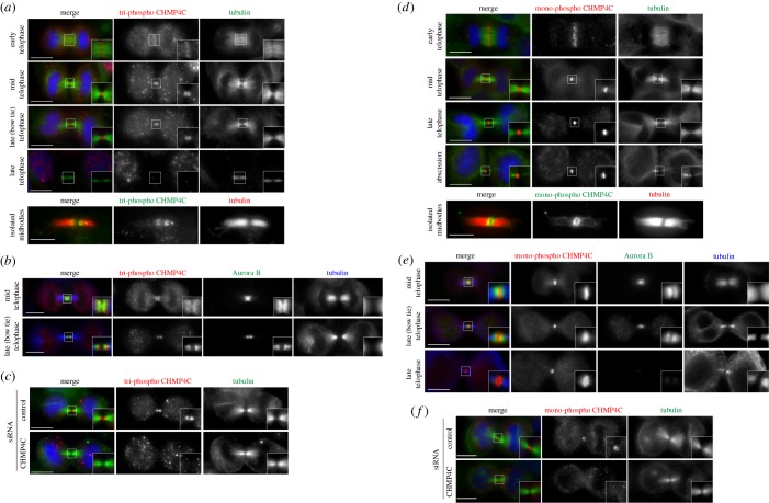
The two phosphorylated forms of CHMP4C display distinct localization patterns during cytokinesis. (a) HeLa Kyoto cells were fixed and stained to detect tri-phospho CHMP4C (red), tubulin (green) and DNA (blue). Insets show two times magnification of the midbody. Scale bars, 10 µm. At the bottom, midbodies were purified form HeLa Kyoto cells and fixed and stained to detect tri-phospho CHMP4C (green) and tubulin (red). Scale bar, 5 µm. (b) HeLa Kyoto cells were fixed and stained to detect tri-phospho CHMP4C (red), Aurora B (green) and tubulin (blue). Insets show two times magnification of the midbody. Scale bars, 10 µm. (c) HeLa Kyoto cells were treated with siRNAs directed against either a random sequence (control) or CHMP4C twice at a 48 h interval and then after 96 h fixed and stained to detect tri-phospho CHMP4C (red), tubulin (green) and DNA (blue). Insets show a two times magnification of the midbody. Scale bars, 10 µm. (d) HeLa Kyoto cells were fixed and stained to detect mono-phospho CHMP4C (red), tubulin (green) and DNA (blue). Insets show two times magnification of the midbody. Scale bars, 10 µm. At the bottom, midbodies were purified form HeLa Kyoto cells and fixed and stained to detect mono-phospho-CHMP4C (green) and tubulin (red). Scale bar, 5 µm. (e) HeLa Kyoto cells were fixed and stained to detect mono-phospho CHMP4C (red), Aurora B (green) and tubulin (blue). Insets show two times magnification of the midbody. Scale bars, 10 µm. (f) HeLa Kyoto cells were treated with siRNAs directed against either a random sequence (control) or CHMP4C twice at a 48 h interval and after 96 h fixed and stained to detect mono-phospho CHMP4C (red), tubulin (green) and DNA (blue). Insets show a two times magnification of the midbody. Scale bars, 10 µm. In all experiments, the shape and thickness of microtubule bundles were used as criteria to stage telophase cells as described [1,3].

