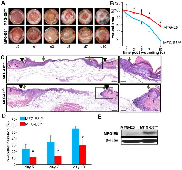
Figure 1. MFG-E8 deficiency impairs wound closure and wound re-epithelialization.
A-D, Full-thickness (skin and panniculus carnosum) dorsal wounds were created on the male MFG-E8−/− and MFG-E8+/+ mice by using a 6-mm biopsy punch. The wounds were stented and left to heal by secondary intention. Wounds were imaged on d0-10 post wounding. Wound areas were calculated using digital planimetry. A, Representative digital images of wounds from age matched MFG-E8−/− and MFG-E8+/+ mice d0-10 post wounding. B, Wound closure kinetics. Data are expressed as mean ± SD (n=5); *p<0.05 compared to MFG-E8+/+ wounds. C, Representative images of hematoxylin-eosin (H&E) stained d7 wound tissues. The wound edge is shown using solid black arrow heads and the wound epithelial margins shown by green arrows. D, Wound re-epithelialization calculated on d5, 7 and 10 post-wounding. Data are expressed as mean ± SD (n=3); *p<0.05 compared to MFG-E8+/+ wounds. E, Western blot analysis of MFG-E8-L protein expression in skin from MFG-E8+/+ and MFG-E8−/− mice. β-Actin was used as a loading control.

