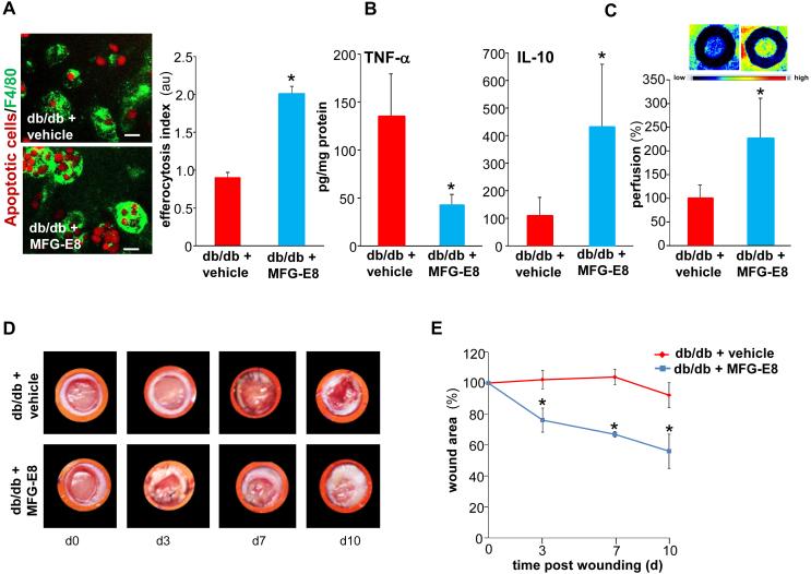
Figure 8. MFG-E8 treatment improves efferocytosis, angiogenesis and healing in diabetic wounds.
A, Efferocytosis activity in d3 wound mϕ harvested from db/db animals. Freshly isolated wound mϕ were incubated with rMFG-E8 (1μg/ml) 2h prior to the subjecting them to efferocytosis in the presence of rMFG-E8 (1μg/ml) or vehicle. Representative images of mϕ (F4/80, green) co-cultured with apoptotic cells (pHrodo, red). Efferocytosis index was calculated and data presented as mean ± SD (n = 4); *p<0.05 compared to vehicle treated group. Scale bar = 10μm. B-E, Full-thickness dorsal wounds were created using a 6-mm biopsy punch on dorsal side of diabetic (Leprdb, db/db) or corresponding non-diabetic controls (heterozygous, Leprdb/+, db/+) mice. The wounds were stented and left to heal by secondary intention. Each wound was either treated with either recombinant mouse MFG-E8 (1μg per wound in 50% glycerol/saline once daily) or equivalent amount of vehicle (mouse serum in 50% glycerine/saline once daily) for 10 days. B, Quantification of TNF-α and IL-10 measured by ELISA in the d10 wound-edge tissues. Data are expressed as mean ± SD (n=3); *p<0.05 compared to vehicle treated wounds. C, Laser Speckle images from d5 wounds. Bar graph presents quantification of the Laser Speckle analysis. Data presented as mean ± SD (n = 4); *p<0.05 compared to control mice. D, Representative wound images on d0-10 post wounding; E, Wound area measurements were done using digital planimetry. Data are expressed as mean ± SD (n=3). *p<0.05 as compared to vehicle treated db/db mice.

