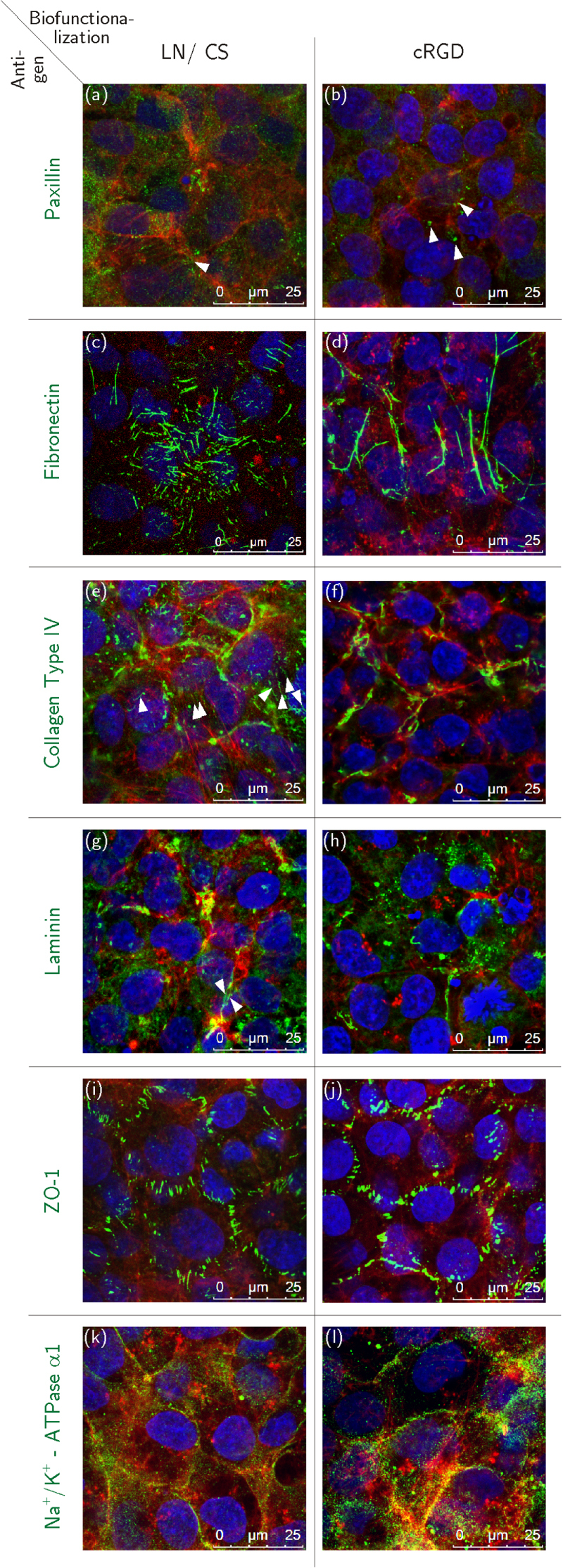
Figure 4.

Immunocytochemical staining of HCEC before detachment; HCEC were cultured for eight days on PVME50–PNiPAAm40–PVMEMA10 carriers functionalized with LN/CS or cRGD. (a), (b) Dot-shaped paxillin signals (white arrowheads). (c), (d) Fibronectin fibers. (e), (f) Fine collagen type IV fibers (white arrowheads) and aggregated collagen type IV. (g), (h) Few fine laminin fibers (white arrowheads) and mostly aggregated laminin signals. (i), (j) ZO-1 localized to the lateral cell membranes. (k), (l) Strong Na+/K+-ATPase α1 signals associated to the lateral cell membranes. Antigens of interest are shown in green (Alexa Fluor®488), F-actin fibers in red (Phalloidin) and the nuclei in blue (Hoechst). n = 3.
