Figure 5.

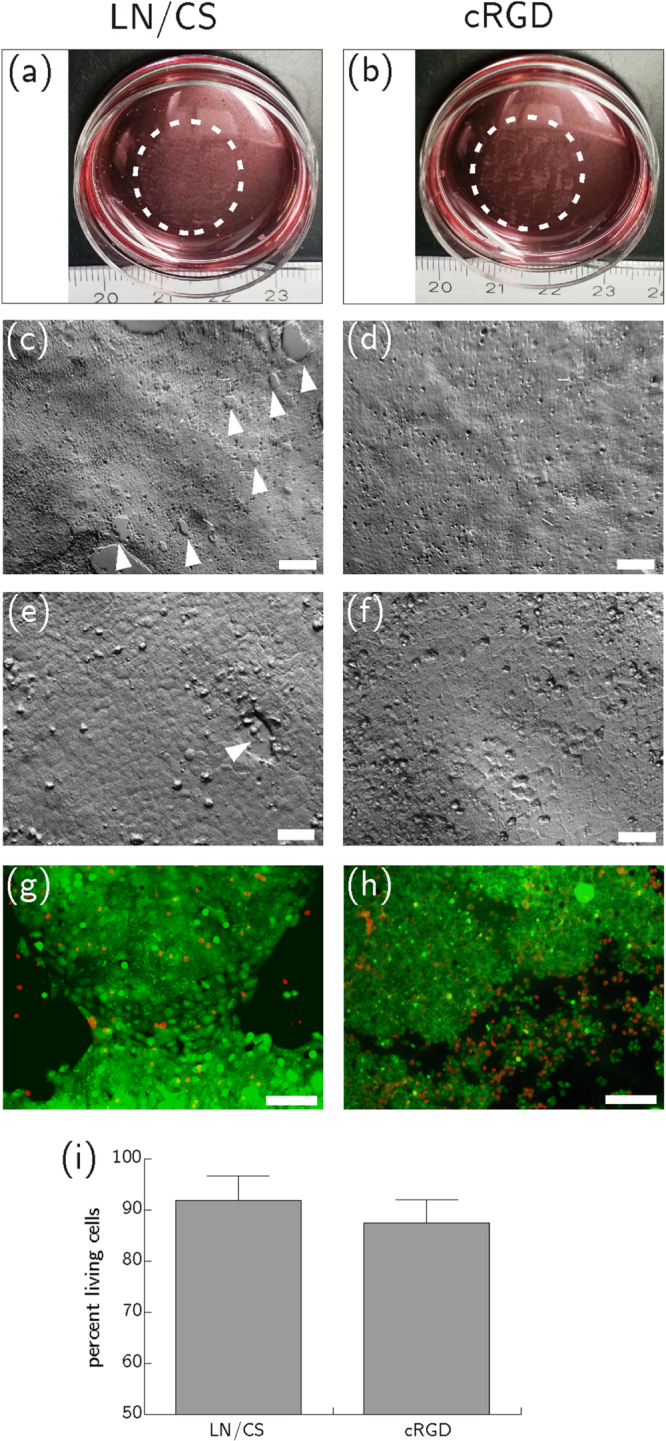
HCEC after thermally induced detachment from PVME50–PNiPAAm40–PVMEMA10 carriers functionalized with LN/CS or cRGD and transfer onto biofunctionalized, polymer-coated cover slips. (a), (b) Macroscopic images show HCEC sheets four hours after the transfer. (c)–(f) Microscopic images of HCEC sheets four hours after the transfer. The few holes that emerged in the fragile cell sheet during detachment and transfer are marked with white arrow heads (scale bar (c) and (d): 200 μm, scale bar (e), (f) 50 μm). (g), (h) Life-dead-staining of HCEC one day after transfer. Vital cells are shown in green (FDA), necrotic cells in red (PI) (scale bar 130 μm). (i) Flow cytometric analysis of HCEC after vital staining with PI. Better cell survival was seen after transfer from LN/CS-functionalized SRP carriers than after transfer from cRGD-functionalized samples. The difference between LN/CS-functionalized and cRGD-functionalized SRP carriers was not statistically significant (p = 0.315). Mean ± sd, n = 3.
