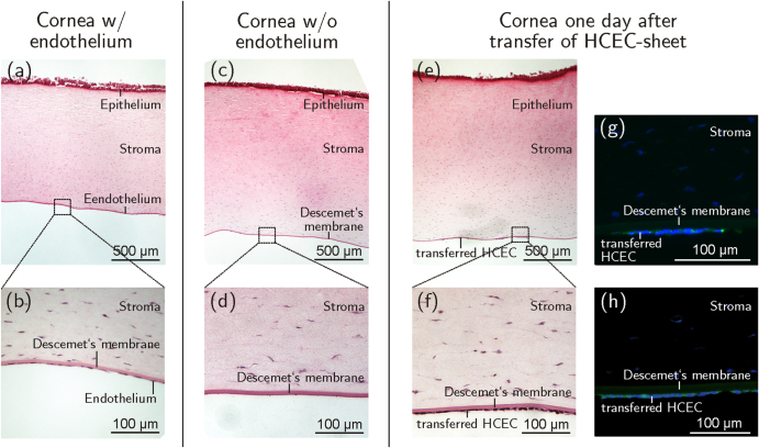
Figure 7.
Sagittal sections of porcine corneas. (a), (b) The corneal endothelium can be seen as a monolayer of squamous cells at the posterior corneal side before de-endothelialization. (c), (d) After removal of the porcine endothelium the bare Descemet’s membrane is visible. (e), (f) One day after transfer of HCEC-sheets onto de-endothelialized corneas the cells appeared attached to Descemet’s membrane as a confluent monolayer. Positive immunohistochemical staining against ZO-1 (g) and Na+/K+-ATPase α1 (h) of transferred HCEC-sheets shown in green (Alexa Fluor®488), nuclei are displayed in blue (DAPI). n = 3.

