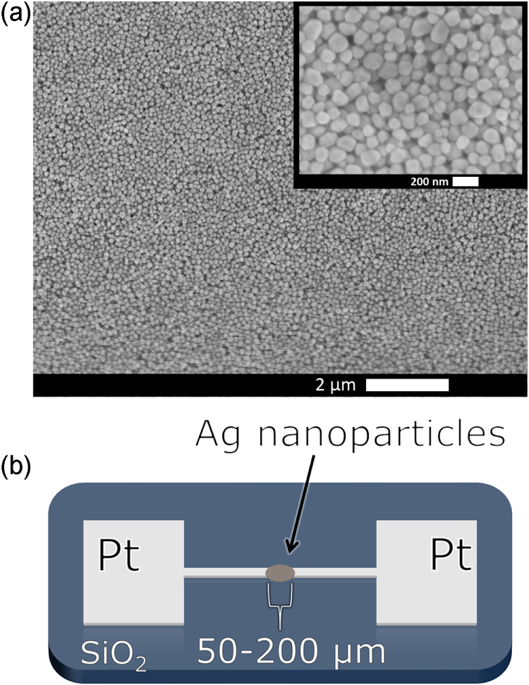
Figure 1.

(a) Scanning electron microscopy image of a silver nanoparticles after synthesis, scale bar = 2 μm. A higher magnification image is inset, scale bar = 200 nm. (b) Schematic of a nanoparticle film device (not drawn to scale). Platinum electrodes with terminals spaced 50–200 μm apart on a SiO2 substrate surround the drop-caste nanoparticles on either side.
