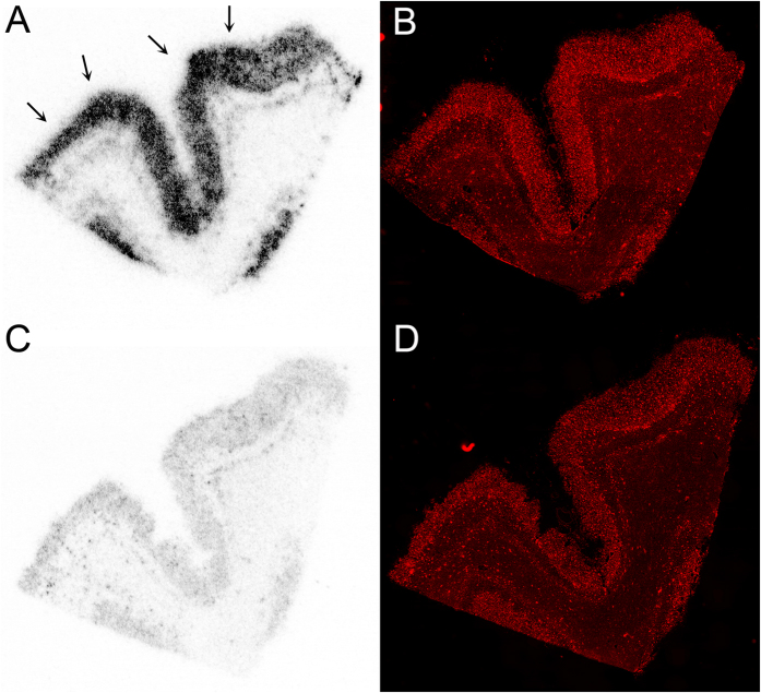
Figure 6.
Autoradiography images of [18F]-9 binding in an AD frontal cortex section following incubation with either [18F]-9 (2 nM) alone (A) or in the presence of 5 (1 μM, C). Fluorescent immunostaining of sections (A) and (C) with an anti-Aβ antibody conjugate is shown in (B) and (D), respectively. The autoradiography images demonstrate laminar distribution of [18F]-9 binding in cortex, which correlates with the distribution of Aβ plaques detected by fluorescent immunostaining, and binding of [18F]-9 is inhibited by excess cold ligand 5 (1 μM, C).

