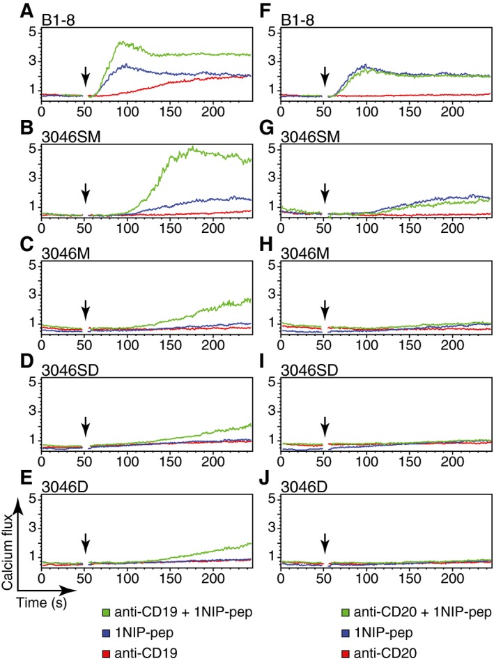
Figure 6. Stimulation of the B cell with anti‐CD19 enhances its response to monovalent antigen.

-
A–EB1‐8 B cells, 3046SM cells, 3046M cells, 3046SD cells or 3046D cells were stimulated with 1NIP‐pep (80 nM), or anti‐CD19 antibody (62 nM), or co‐stimulated with 1NIP‐pep and anti‐CD19 antibody. The calcium fluxes were measured by FACScan.
-
F–JB1‐8 B cells, 3046SM cells, 3046M cells, 3046SD cells or 3046D cells were stimulated with 1NIP‐pep (80 nM), or anti‐CD20 antibody (62 nM), or co‐stimulated with 1NIP‐pep and anti‐CD20 antibody. The calcium fluxes were measured by FACScan.
