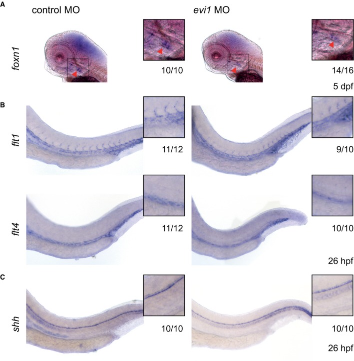
WISH of foxn1 in the thymus epithelium of both control (left)‐ and evi1 MO‐injected (right) 5 dpf embryos.
WISH of endothelial‐specific flt1 (upper) and flt4 (lower) in both control (left)‐ and evi1 MO‐injected (right) 26 hpf embryos.
WISH of shh in the notochord of both control‐ and evi1 MO‐injected embryos.
Data information: Lateral views are shown, with anterior to the left, dorsal up. Squares represent enlargements of the region of interest. Numbers indicate the amount of embryos with the respective phenotype/total number of embryos analyzed in each experiment. A minimum of two biological replicates was performed for each marker with at least
n = 5 embryos per experiment.

