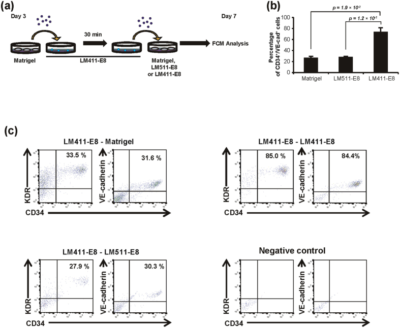
Figure 3. Effects of sequential switching of matrices during endothelial differentiation.
(a) Schematic process of the double-switching assay. Day 3 cells were dissociated at the single-cell level and plated onto LM411-E8-coated plates. After 30 minutes of incubation at 37 °C, adherent cells were replated onto Matrigel, LM511-E8 and LM411-E8 and cultured with VEGF stimulation for four days. (b) Percentage of PSC-EPCs (KhES-1) differentiated from LM411-E8-selected mesodermal progenitors on Matrigel, LM511-E8 or LM411-E8 for four days. Data are presented as the mean ± SEM (n = 3) and were statistically analyzed using Student’s t-test. Representative results of at least two independent experiments are shown.

