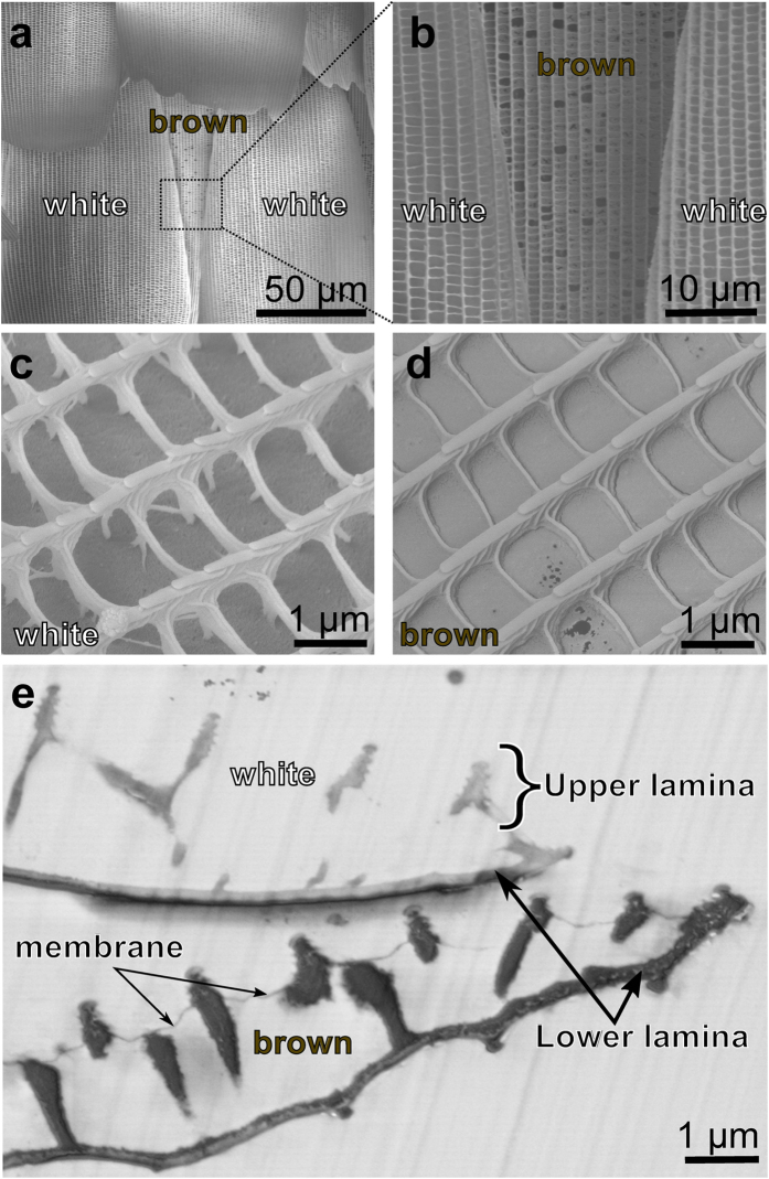
Figure 2. Micro- and nanostructure of the Hypolimnas salmacis scales.
(a,b) SEM images of stacks of white cover scales on brown ground scales. (c) Detail of a single white scale. Longitudinal grating like ridges along the scale are connected with cross-ribs. The sides of the ridges are covered with small microribs. (d) Detail of single brown scale. In terms of dimension, it mimics almost exactly the white scale but the windows, created by the cross-ribs, are closed by thin pigmented membranes. (e) SEM image of a cross section of the forewing including a white and brown scale. Both scales show similar features with upper lamina of ridges and tiny microribs as well as lower lamina of thin films. Thin membranes between the ridges are visible only in the brown scale.

