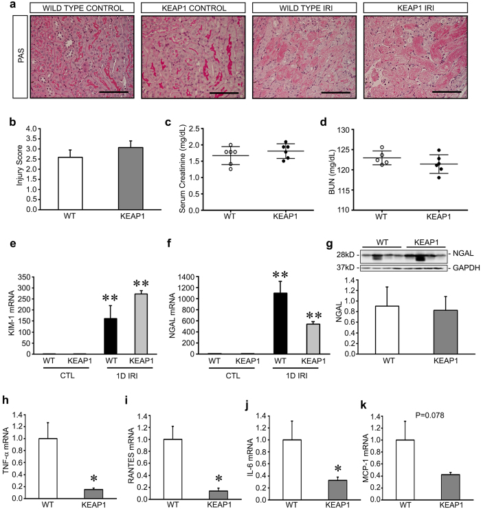
Figure 1. Keap1 hypomorphs demonstrate minimal protection 24 hours after ischemia-reperfusion injury (IRI).
Keap1 hypomorphs (KEAP1) and wild type (WT) controls were subjected to unilateral renal IRI with simultaneous contralateral nephrectomy. (a) Histological assessment with Periodic Acid Schiff (PAS) staining showed similar tubular damage after injury, as manifested by tubular necrosis and loss of brush borders, in both sets of mice compared to the untreated control kidney (which themselves were not different between groups). Bar equals 100 μm. (b) Quantitative analysis confirmed no detectable difference in histologic injury (n = 6 for each group). (c,d) There were no significant differences in serum creatinine and BUN between groups. Each dot represents an individual animal with the mean ± SEM superimposed. (e,f) After IRI, qRT-PCR analysis of tubular injury markers KIM-1 and NGAL revealed significant increases from the control kidneys, but no difference between WT and KEAP mice. (**P < 0.05 compared to either CTL untreated kidney values, one-way ANOVA, n = 4). (g) Western blot analysis and densitometry of NGAL confirms no significant difference between injured WT and KEAP1 kidneys. NGAL was not detectable by this method in uninjured kidneys. (h–k) Proinflammatory mediators were reduced in KEAP1 compared to WT mice (n = 5–6 in each group). (*P < 0.05 compared to WT mice, P = 0.078 for MCP-1.)

