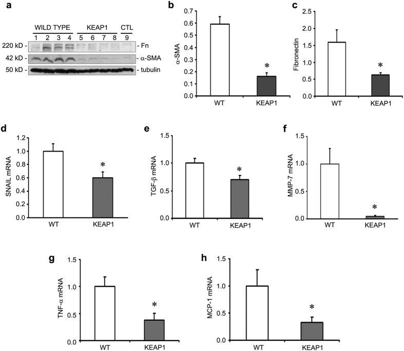
Figure 4. Keap1 hypomorphs were protected from the fibrotic and inflammatory response at 10 days after ischemia-reperfusion injury (IRI).
(a) Western blot for fibrosis markers fibronectin (Fn) and smooth muscle actin (α-SMA) along with the loading control tubulin is shown for IRI wild type and KEAP1 mice, along with an untreated control (CTL) for reference. (b,c) Western blot densitometry of Fn and α-SMA (normalized to tubulin) shows significant reduction in the hypomorphs. (d–f) qRT-PCR of fibrosis-related genes Snail, MMP-7, and TGF-β (and α-SMA, data not shown) were significantly reduced in Keap1 hypomorphs. (n = 5–6 for each group) (g,h) Hypomorphs were also protected from increases in mRNA for proinflammatory cytokines TNF-α and MCP-1. (*P < 0.05 compared to the wild type mice. N = 5–6).

