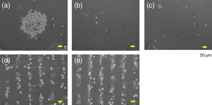
Figure 7.

Patterning of NIH3T3 cells on the substrates in a serum-containing medium. (a–c) Cell adhesion to the 70% EG7 substrate (a) with or (b) without His-FNIII7–10 (50 μg ml-1) or (c) with pFN (50 μg ml-1). The substrates were irradiated in a circular pattern. (d, e) Cell adhesion on the 50% EG7 substrate (d) with or (e) without His-FNIII7–10 (50 μg ml-1). The substrates were irradiated in a striped pattern. Cells were allowed to attach for 4 h and phase-contrast images were obtained after removing the unattached cells. The %NTA was 5% for (a–c) and 0.5% for (d) and (e). Scale bars represent 50 μm.
