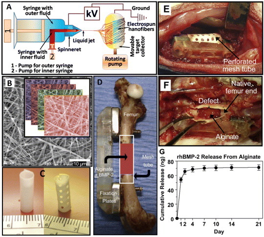
Figure 8.

(A) Schematic of a coaxial electrospinning setup. (B) Scanning electron microscopy image of electrospun nanofiber mesh illustrating the smooth and bead-free fibers with nanosized diameters; the inset shows the layers of fibers in a scaffold. (C) Hollow tubular implant made from nanofiber meshes without and with perforations. (D) In vivo application of the nanofiber mesh tubes as implants placed around an 8 mm segmental femoral rat bone defect (in some groups, alginate hydrogel, with or without rhBMP-2, is injected inside the hollow tube). (E) Defect after implantation of a perforated mesh tube; the alginate inside the tube can be seen through the perforations. (F) After 1 week, the mesh tube was cut open, and the alginate was still present inside the defect with hematoma at the bone ends. (G) In vitro alginate release kinetics: sustained release of the rhBMP-2 was observed during the first week (images B-G are taken from [97] with permission).
