
Table 1.
Properties of TE scaffolds desired for different tissues and effects of electrospun materials on TE scaffolds.
| Tissue | Biological description | Key engineering properties | Effect of electrospun materials on TE scaffolds | Use of scaffolds |
|---|---|---|---|---|
| Epithelial tissue e.g. skin | Joined together with same tissue; soft and elastic | Very low elastic moduli (0.1–0.2 MPa), optimum pore size (20–125 μm) [22] | Electrospun collagen nanofibers can improve the structural integrity and mechanical strength of skin tissues. The scaffolds made of electrospun nanofibers provide a high surface area-to-volume ratio, which promotes the cell–matrix interaction at the nanoscale [23] | 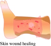 |
| Connective tissue e.g. bone, tendon, ligament, cartilage and fat | Joins different tissues; strong and tough | High tensile, compressive, and torsional strengths, high elastic moduli, optimum pore size for bone (100– 250 μm) [24], ideal porosity (>90%) [25] | Electrospun nanofibers with high surface porosity improve cell ingrowth and the mechanical properties of the scaffold [26,27] | 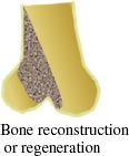 |
| Fluid tissue e.g. blood, fibrinogen (natural polymer present in blood plasma), thrombin | Transports food, nutrients and waste products; viscous | Specified viscosity, surface tension, mass transport property and pH | The high surface-to-volume ratio of the electrospun synthetic fibrinogen nanofibers can improve the blood clotting process in wound healing after reacting with thrombin by forming a network structure of a fibrous compound called fibrin [28] | 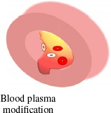 |
| Nerve tissue | Sensitive to various stimuli | Smart properties; high ionic, electrical and thermal conductivities, electrochemical and chemoelectrical transduction properties [29] | High aspect ratio of electrospun nanofibers enhances the conductivity of sensors via electron transport, which is extremely effective for nerve tissue scaffolds [29] | 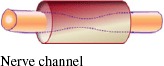 |
| Muscle tissues: | ||||
| Voluntary muscle tissue e.g. arm, leg and skeletal muscles | Made of striated muscle fibers supported by connective tissues and stimulated by nerves | Medium elastic modulus and high fatigue endurance under cyclic load | Electrospun nanofibers of polyester urethane and poly(l-lactide-co-ε-caprolactone) have satisfactory mechanical properties and encouraging cellular response in terms of adhesion and differentiation; they can be used in scaffolds for skeletal [30] or smooth [31] muscles |  |
| Involuntary muscle tissue e.g. intestines, heart or cardiac muscles | Smooth and not human-controlled; soft | Low elastic modulus and high fatigue endurance limit under cyclic load | Small intestinal submucosa (SIS) composed of type-I and type-III collagens and various cytokines leads to superior initial cell attachment and proliferation compared with synthetic polymeric scaffolds in presence of growth factors. Electrospun SIS/poly(ε-caprolactone) hybrids have a stable micro/nanofibrous structure, which provide improved hydrophilicity, mechanical properties and cellular behavior to the scaffolds [32] |  |