
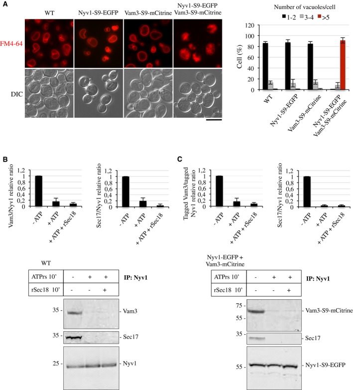
Figure 5. Effect of tags on vacuole morphology and cis‐SNARE activation.
-
AIn vivo morphology. Cells expressing the indicated SNARE variants were grown in SC medium (16 h, 30°C), stained with FM4‐64, and their relative morphology was assessed by fluorescence and DIC microscopy. Scale bar: 5 μm. The cells were grouped into three categories according to the number of vacuoles visible per 100 cells. Values represent the means and s.d. from three independent experiments.
-
B, CSNARE activation on isolated vacuoles. Vacuoles were isolated from a strain co‐expressing NYV1‐S9‐EGFP and VAM3‐S9‐mCitrine and from an isogenic wild type. One hundred and fifty micrograms of the organelles was incubated in fusion reactions in the presence or absence of an ATP‐regenerating system and recombinant, purified Sec18/NSF (rSec18, 50 μg/ml). After 10 min of incubation at 27°C, the vacuoles were solubilized and immunoprecipitated with antibodies to Nyv1. Co‐immunoprecipitated proteins were analyzed by SDS–PAGE and Western blotting. The histograms provide quantifications of the band intensities as the means ± s.d. from three independent experiments.