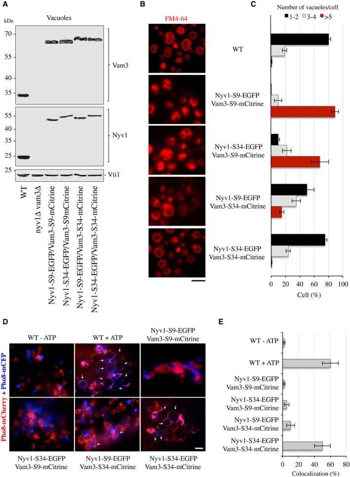
Vam3 and Nyv1 were tagged with mCitrine and EGFP as shown Fig
3A, but the spacer was extended (S34) by an additional 25 amino acid sequence (SGGGGSGGGGSGGGGSGGGGAAAGG).
Protein levels on isolated vacuoles from these strains were compared as in Fig
3B.
Vacuole morphology was assessed as in Fig
3C. Scale bar: 5 μm.
The cells were grouped into three categories according to the number of vacuoles visible per 100 cells. Values represent the means and s.d. from three independent experiments.
Fusion activity: Vacuoles were isolated from BJ3505 strains expressing the indicated versions of Vam3 and Nyv1 and Pho8‐mCFP or Pho8‐mCherry. Ten micrograms of vacuoles was incubated in standard fusion reactions in the presence or absence of ATP and analyzed by confocal microscopy. Arrows indicate examples of fusion products. Scale bar: 5 μm.
Vacuole fusion was assayed by measuring the percentage of colocalization of the Pho8‐mCFP and Pho8‐mCherry signals. Means ± s.d. are shown for at least 100 vacuoles from three experiments.

